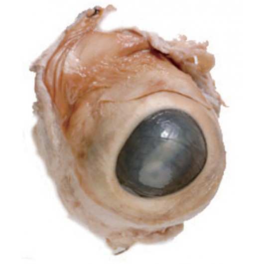
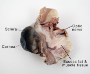
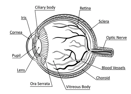
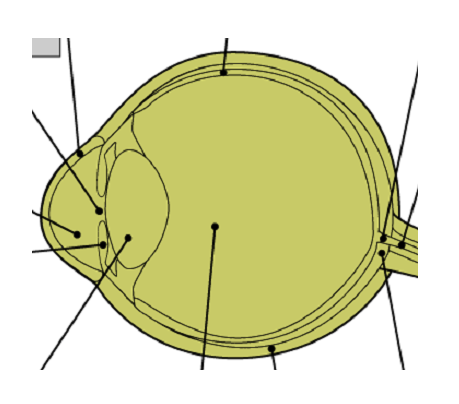
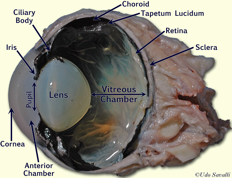
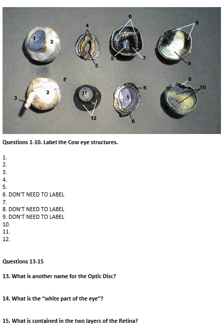
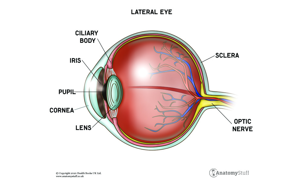
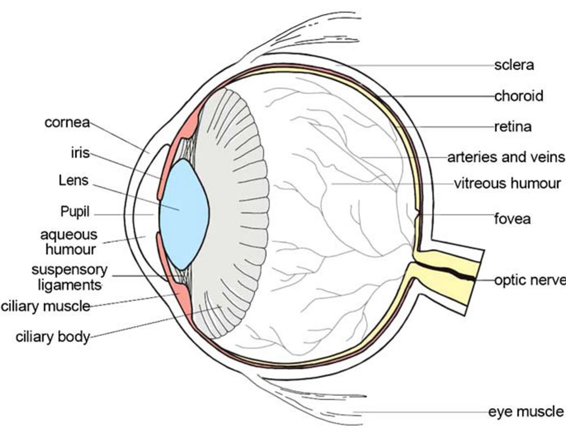
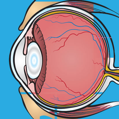
42 cows eye labeled
13.7: Cow Eye Dissection - Biology LibreTexts When the cow was alive, the cornea was clear. In your cow's eye, the cornea may be cloudy or blue in color. 2. Cut away the fat and muscle, this may only be necessary if fat is covering the cornea of the eye and is in your way. Fat around the backside of the eye can be left alone. Cow's Eye Dissection - Eye diagram - Exploratorium Learn how to dissect a cow's eye in your classroom. This resource includes: a step-by-step, hints and tips, a cow eye primer, and a glossary of terms. Cow's Eye Dissection - Eye diagram
Cow Eye Dissection - YouTube Apr 26, 2013 ... Cow Eye Dissection. Watch later. Share. Copy link. Info. Shopping. Tap to unmute. If playback doesn't begin shortly, try restarting your ...

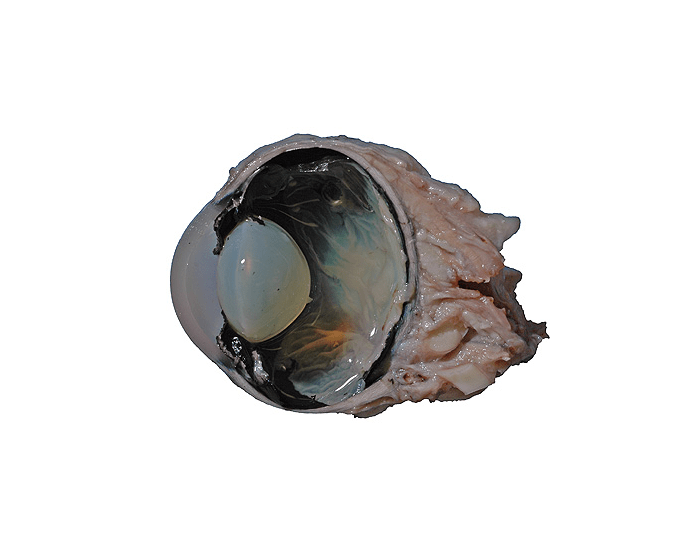
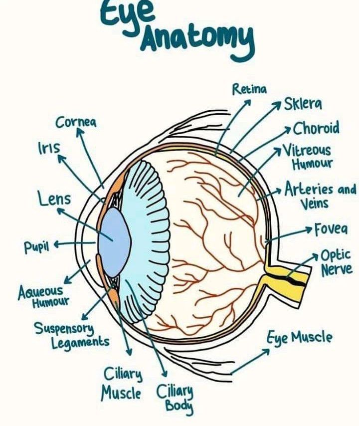
Cows eye labeled
PDF Table 8.1: External Anatomy of The Cow Eye Feature Description TABLE 8.2 CONTINUED: INTERNAL ANATOMY OF THE COW EYE FEATURE DESCRIPTION Retina Innermost layer of the eye; only in posterior cavity; delicate, thin, cream colored sheet of tissue Optic disc A single point of attachment of the retina - to the optic nerve (also called the blind spot) Choroid Middle layer of the eye, posterior portion; 10 Cow eye ideas - Pinterest See more ideas about cow eyes, eye anatomy, anatomy and physiology. ... Cow's Eye Dissection - Eye diagram Human Eye Diagram, Diagram Of The Eye, Veterinary. anatomy cow eye quiz Flashcards | Quizlet iris. what part of the cow eye is this. Cornea. what is this part of the cow eye. retina. what is this part of the cow eye. tapetum lucidum. what part of the cow eye is the red arrow pointing to. nerve endings in muscle.
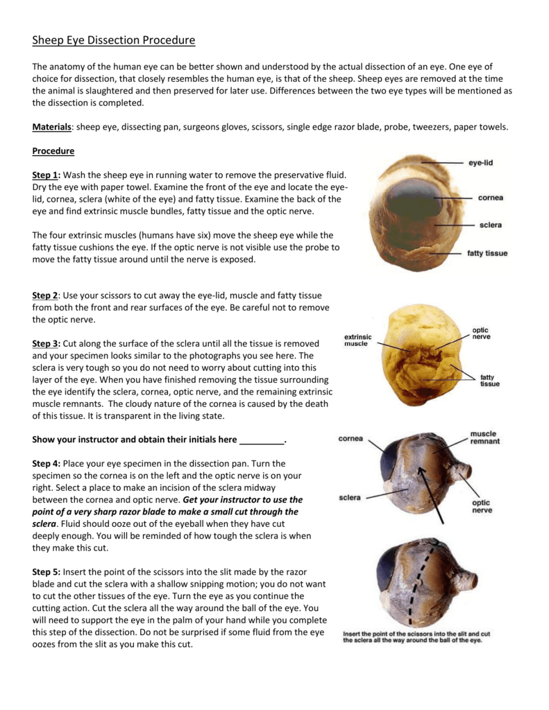
Cows eye labeled. Cow Eye Dissection & Anatomy Project | HST Learning Center Look carefully at the preserved cow eye. The most noticeable part of the eye is the large mass of gray tissue that surrounds the posterior(back) of the eye and is attached to the sclera. The second most noticeable part of the eye is the cornea, located in the anterior(front) part of the eye. Cow Eye Dissection & Labeling - YouTube May 24, 2018 ... Cow Eye Dissection & Labeling. 48K views 4 years ago. Brentt J. Swetter. Brentt J. Swetter. 56 subscribers. Subscribe. Cow's Eye Dissection | Exploratorium Learn how to dissect a cow's eye in your classroom. This resource includes: a step-by-step, hints and tips, a cow eye primer, and a glossary of terms. PDF COW'S EYE dissection - Exploratorium When the cow was alive, the cornea was clear. In your cow's eye, the cornea may be cloudy. You may be able to look through the cornea and see the iris, the colored part of the eye, and the pupil, the dark oval in the middle of the iris. Cut away the fat and muscle. Use a scalpel to make an incision in the cornea. (Careful—Don't cut yourself!)
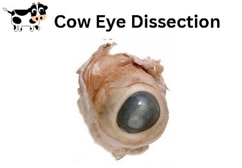
Cow Eye Dissection Guide - Google Slides Cow Eye Many laboratory books label the fovea centralis/macula, which is the location in eye where the sharpest vision occurs; the fovea centralis/macula is dense with cones and is the... Cow Eye Dissection | Carolina.com Cow Eye Internal Anatomy Hold the eye between your thumb and forefinger, as shown below. Using scissors or a scalpel, carefully cut the eye in half, separating the front and back of the eye. Examine the inside front portion of the eye. Remove the gelatinous vitreous humor and hard lens. Cow's Eye Dissection - step 1 - Exploratorium Here's a cow's eye from the meat company. The white part is the sclera , the outer covering of the eyeball. The blue is the cornea , which starts out clear but becomes cloudy after death. WATCH VIDEO © Exploratorium | The museum of science, art and human perception The thick, tough, white outer covering of the eyeball. Cow Eye Dissection - Biology LibreTexts 1. Examine the outside of the eye. You should be able to find the sclera, or the whites of the eye. This tough, outer covering of the eyeball has fat and muscle attached to it. 2. Locate the covering over the front of the eye, the cornea. When the cow was alive, the cornea was clear. In your cow's eye, the cornea may be cloudy or blue in color.
Cow Eye Dissection - The Biology Corner 1. Examine the outside of the eye. You should be able to find the sclera, or the whites of the eye. This tough, outer covering of the eyeball has fat and muscle attached to it. 2. Locate the covering over the front of the eye, the cornea. When the cow was alive, the cornea was clear. In your cow's eye, the cornea may be cloudy or blue in color. Cow's Eye Dissection | Exploratorium Step 1: The cow's eye Here's a cow's eye from the meat company. The white part is the sclera, the outer covering of the eyeball. The blue is the cornea, which starts out clear but becomes cloudy after death. Step 2: Muscles move the eye Without moving your head, look up. Look down. Look all around. Dissecting An Eyeball | Lyncean Education Cow eyes are also readily available, as a byproduct of beef production, and can be purchased inexpensively from science supply companies, such as Carolina Biological Supply. Such eyeballs are normally stored in preservative, which has the unfortunate side effect of making the cornea and lens cloudy. Cow Eye Anatomy Flashcards | Quizlet Cornea Clear protective covering over the front of eye that bends light entering eye Pupil The hole where light passes into the lens Iris The colored part of the eye that regulates the size of the pupil to control light entering the eye Vitreous Humor A clear liquid inside eyeball that gives the eye its round shape Lens
Detailed Cow Eye Dissection: Part I (Jr. High, High ... - YouTube Feb 4, 2014 ... Cow Eye dissection for educational use: additional video, lesson plans, quizzes, additional dissections and more available at ...
Cow Eye Labeling Quiz - PurposeGames.com Cow Eye Labeling by secretsquirrel 21,896 plays 11 questions ~30 sec English 11p More 24 4.83 (you: not rated) Tries Unlimited [?] Last Played March 1, 2023 - 03:54 am There is a printable worksheet available for download here so you can take the quiz with pen and paper. Remaining 0 Correct 0 Wrong 0 Press play! 0% 10:00.0 Show More
Cow eye - dissection and label - SlideShare Cow eye shown with labeled cornea. The cornea is the transparent front part of the eye that covers the iris, pupil, and anterior chamber. The cornea, with the anterior chamber and lens, refracts light, with the cornea accounting for approximately two-thirds of the eye's total optical power.
Ultrasonographic anatomy of the bovine eye - PubMed Transpalpebral ultrasonographic images were obtained with a 10 MHz linear transducer in both horizontal and vertical imaging planes. The ultrasonographic appearance of structures within the bovine eye is similar to that in other species, although the ciliary artery was frequently identified, appearing as a 0.33 +/- 0.04 cm diameter hypoechoic ...
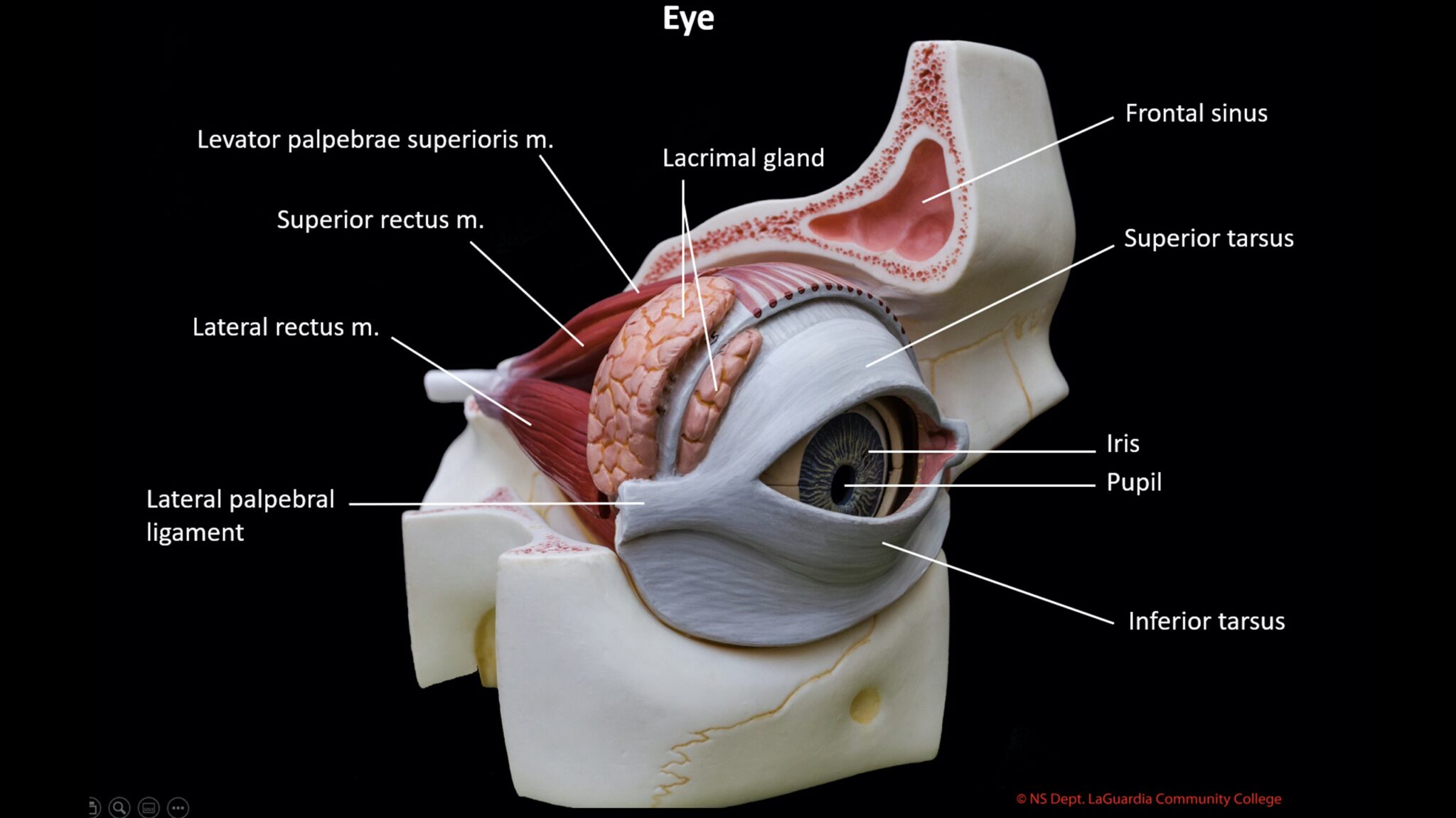
What are the Differences Between a Cow Eye & Human Eye? The eyeballs of humans and the eyeballs of cows have a similar structure overall. Both have the sclera, which is the white part of the eyeball, cornea or the clear structure over the iris and pupil, lens, vitreous fluid, retina and choroid. The choroid is the layer of the eyeball that is between the retina and the sclera.
Cow Eye Dissection & Parts of the Eye Diagram | Quizlet Photoreceptor cells in the eye that detect black, white, and gray cones Photoreceptor cells in the eye that detect color aqueous humor Fluid in front chamber of eye; nourishes the cornea and the lens. vitreous humor The clear gel filling the space of the eyeball between the lens and the retina macula
anatomy cow eye quiz Flashcards | Quizlet iris. what part of the cow eye is this. Cornea. what is this part of the cow eye. retina. what is this part of the cow eye. tapetum lucidum. what part of the cow eye is the red arrow pointing to. nerve endings in muscle.
10 Cow eye ideas - Pinterest See more ideas about cow eyes, eye anatomy, anatomy and physiology. ... Cow's Eye Dissection - Eye diagram Human Eye Diagram, Diagram Of The Eye, Veterinary.
PDF Table 8.1: External Anatomy of The Cow Eye Feature Description TABLE 8.2 CONTINUED: INTERNAL ANATOMY OF THE COW EYE FEATURE DESCRIPTION Retina Innermost layer of the eye; only in posterior cavity; delicate, thin, cream colored sheet of tissue Optic disc A single point of attachment of the retina - to the optic nerve (also called the blind spot) Choroid Middle layer of the eye, posterior portion;













Komentar
Posting Komentar