45 compound microscope labeled
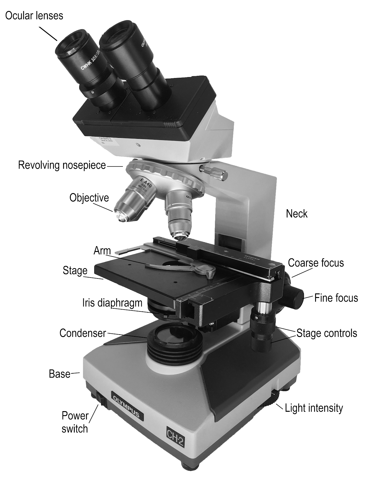
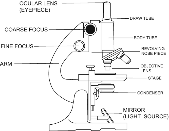
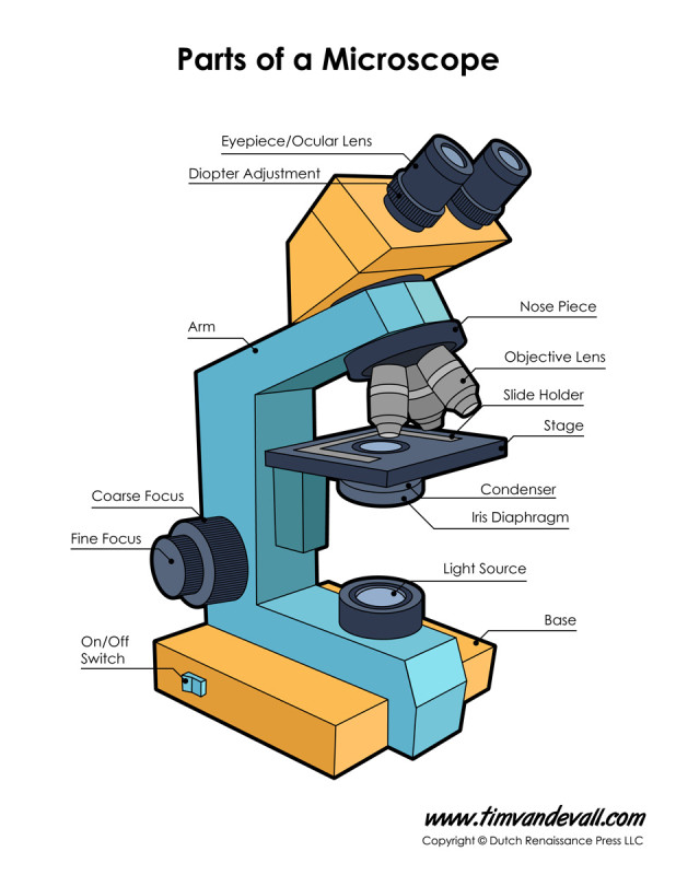
Compound Microscope Parts, Functions, and Labeled Diagram Compound Microscope Definitions for Labels. Eyepiece (ocular lens) with or without Pointer: The part that is looked through at the top of the compound microscope. Eyepieces typically have a magnification between 5x & 30x. Monocular or Binocular Head: Structural support that holds & connects the eyepieces to the objective lenses. Microscope Parts and Functions With Labeled Diagram and Functions How does a Compound Microscope Work? Before exploring microscope parts and functions, you should probably understand that the compound light microscope is more complicated than just a microscope with more than one lens. Parts of a Chainsaw - Anatomy Explained [2022 Update]
UD Virtual Compound Microscope - University of Delaware ©University of Delaware. This work is licensed under a Creative Commons Attribution-NonCommercial-NoDerivs 2.5 License.Creative Commons Attribution-NonCommercial-NoDerivs 2.5 License.

Compound microscope labeled
Imaging Subcellular Structures in the Living Zebrafish Embryo The relative ease with which these subcellular structures can be genetically labeled by fluorescent proteins and the use of light microscopy techniques to image them is ... it is also a fluorescent compound itself. Therefore, Phenol Red could obscure visualizing FPs if imaging is planned before 24 hr post-fertilization (hpf) as it may not be ... Compound Microscope Labeled Diagram | Quizlet Compound Microscope Labeled + − Flashcards Learn Test Match Created by meganplocher734 Terms in this set (14) Eyepiece/Ocular lens Contains the ocular lens Body tube A hollow cylinder that holds the eyepiece. Arm Part that supports the microscope. Stage Supports the slide or specimen Coarse adjustment Knob Compound Microscope: Definition, Diagram, Parts, Uses, Working ... - BYJUS A compound microscope is defined as A microscope with a high resolution and uses two sets of lenses providing a 2-dimensional image of the sample. The term compound refers to the usage of more than one lens in the microscope. Also, the compound microscope is one of the types of optical microscopes.
Compound microscope labeled. What is a Stereo Microscope? - New York Microscope Company 11.05.2018 · Each part of a stereo microscope is labeled in the diagram below. This example is a typical classroom type stereo microscope with track stand and built-in illumination. Stereo Head: This is the moveable top portion of the microscope and the stereo head holds the two adjustable eyepieces. Ocular Lens: These are the eyepieces through which the viewer looks at the … Compound Microscope: Parts of Compound Microscope - BYJUS (A) Mechanical Parts of a Compound Microscope 1. Foot or base It is a U-shaped structure and supports the entire weight of the compound microscope. 2. Pillar It is a vertical projection. This stands by resting on the base and supports the stage. 3. Arm The entire microscope is handled by a strong and curved structure known as the arm. 4. Stage Microscope Types (with labeled diagrams) and Functions A compound microscope: Is used to view samples that are not visible to the naked eye Uses two types of lenses - Objective and ocular lenses Has a higher level of magnification - Typically up to 2000x Is used in hospitals and forensic labs by scientists, biologists and researchers to study micro organisms Compound microscope labeled diagram Microscopy Pre-lab Activities - University of Delaware Microscope controls: turn knobs (click and hold on upper or lower portion of knob) throw switches (click and drag) turn dials (click and drag) move levers (click and drag) changes lenses (click and drag on objective housing) select a specimen (click on a slide)
Compound Microscope Parts - Labeled Diagram and their Functions The term "compound" refers to the microscope having more than one lens. Basically, compound microscopes generate magnified images through an aligned pair of the objective lens and the ocular lens. In contrast, "simple microscopes" have only one convex lens and function more like glass magnifiers. Fluorescence Microscopy- Definition, Principle, Parts, Uses 12.04.2022 · Buy Fluorescence Microscope from LEAM Solution Inc. Typical components of a fluorescence microscope are: Fluorescent dyes (Fluorophore) A fluorophore is a fluorescent chemical compound that can re-emit light upon light excitation. Fluorophores typically contain several combined aromatic groups, or plane or cyclic molecules with several π bonds. EOF Fluorescence Labeling of Neurotensin(8-13) via Arginine Residues Gives ... The indolinium-type cyanine dye-labeled NT(8-13) derivatives 6, 9, 11, 12, 16, and 17 were prepared by treatment of the amino-functionalized precursor peptides 4, 7, or 15, 19 containing an N ω-carbamoylated arginine either in position 8 (4, 7) or in position 9 (15) with the succinimidyl esters of the respective dyes (5, 8, or 10) (Scheme 1).The pyridinium dye-labeled peptide 14 was ...
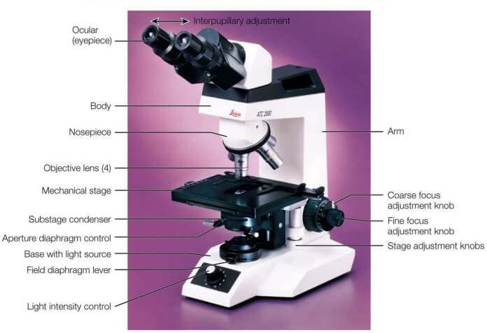
Parts of a Compound Microscope - Labeled (with diagrams) A compound microscope is known as a high-power microscope that enables you to achieve a high level of magnification. Smaller specimens can be thoroughly viewed using a compound microscope. ... Image 3: A compound microscope with a corresponding label of the different parts. imagesource: images.slideplayer.com. The optical components of a ... Compound Light Microscope: Everything You Need to Know A compound microscope is a type of light microscope that uses a compound lens system to magnify specimens for up to 1000x or more. It's made up of at least one objective lens and at least one ocular lens, as well as a light source, condenser, and other essential parts. compound microscope parts (labeling) Flashcards | Quizlet compound microscope parts (labeling) Flashcards | Quizlet compound microscope parts (labeling) Term 1 / 14 eyepiece tube - connects the eyepiece to the objective lens Click the card to flip 👆 Definition 1 / 14 what is 1? Click the card to flip 👆 Flashcards Learn Test Match Created by barnettlily Terms in this set (14) How to Calculate the Magnification of a Microscope? 07.06.2021 · For instance, if the eyepiece is labeled as 30x/18, then 18 ÷ 30 = 0.6, meaning that the diameter of for is 0.6 millimeters. For microscopes that only have an eyepiece, this will be enough. But having an objective lens as well makes the calculation harder. You must multiply the eyepiece magnification with the objective magnification and then divide the field number. If the …
Microscope Labeling
Fluorescence Microscope: Principle, Types, Applications 18.05.2020 · The filters are often plugged together in a filter cube (compound microscopes) or a flat holder (mainly stereo microscopes). Principle. To observe the sample through a fluorescence microscope, it should first be labeled with fluorescent dyes/substances known as fluorophores. Working mechanism of Fluorescence Microscope (Photo credit: Henry Mühlpfordt) Higher …

Activity 1: Name Me!Directions: Identify the parts of the ...
Compound Light Microscope Labelling Quiz - PurposeGames.com This is an online quiz called Compound Light Microscope Labelling. There is a printable worksheet available for download here so you can take the quiz with pen and paper. Your Skills & Rank. Total Points. 0. Get started! Today's Rank--0. Today 's Points. One of us! Game Points. 15.
9.1: Using Microscopes - Biology LibreTexts
Compound Microscope - Diagram (Parts labelled), Principle and Uses Why is it called compound microscope? Because it has multiple lenses that work in conjunction to magnify a specimen Q 4. What are the 13 parts of a microscope? 1. Eyepiece 2. Eyepiece Tube 3. Objective Lens 4. Stage 5. Stage Clips 6. Nosepiece 7. Fine and Coarse Focus knobs 8. Illuminator 9. Aperture 10. Iris Diaphragm 11. Condenser 12.
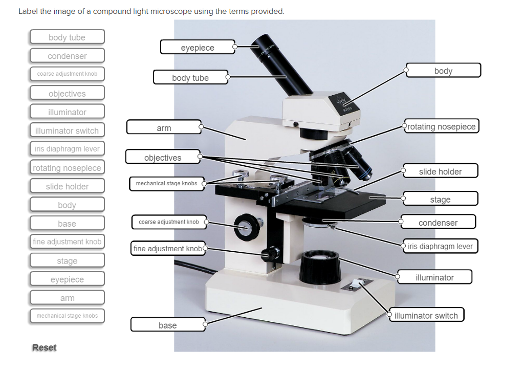
Solved Label the image of a compound light microscope using ...
Parts of a microscope with functions and labeled diagram 17.09.2022 · Q. Differentiate between a condenser and an Abbe condenser. Ans. Condensers are lenses that are used to collect and focus light from the illuminator into the specimen. They are found under the stage next to the diaphragm of the microscope. They play a major role in ensuring clear sharp images are produced with a high magnification of 400X and above.

Difference between Simple and Compound Microscope ...
Parts of Stereo Microscope (Dissecting microscope) – labeled … If you would like to learn optical components of a compound microscope, please visit Compound Microscope Parts – Labeled Diagram and their Functions, and this article. How to use a stereo (dissecting) microscope. Follow these steps to put your stereo microscopes in work: 1. Set your microscope on a tabletop or other flat sturdy surface where ...

Solved Microscope parts/labeling 9 Label the image of a ...
Labelled Diagram of Compound Microscope The below mentioned article provides a labelled diagram of compound microscope. Part # 1. The Stand: The stand is made up of a heavy foot which carries a curved inclinable limb or arm bearing the body tube. The foot is generally horse shoe-shaped structure (Fig. 2) which rests on table top or any other surface on which the microscope in kept.

Can someone can send me diagram of this compound microscope ...
September 26 - Today in Science History - Scientists born on September ... By 1866, their company was making a simple microscope. The company name was changed to Bausch & Lomb Optical Co. in 1874, the year they produced their first compound microscope. Edward, with brothers William, and Henry all helped in the design and production of a full product line of microscopes. ... labeled 'K' and 'L'. In 1916, Siegbahn ...

easy compound microscope diagram - Clip Art Library
Compound Microscope- Definition, Labeled Diagram, Principle, … A compound microscope is of great use in pathology labs so as to identify diseases. Various crime cases are detected and solved by drawing out human cells and examining them under the microscope in forensic laboratories. The presence or absence of minerals and the presence of metals can be identified using compound microscopes.

Labeling the Parts of the Microscope | Microscope World Resources
A Study of the Microscope and its Functions With a Labeled Diagram ... To better understand the structure and function of a microscope, we need to take a look at the labeled microscope diagrams of the compound and electron microscope. These diagrams clearly explain the functioning of the microscopes along with their respective parts. Man's curiosity has led to great inventions. The microscope is one of them.

Living Environment Course
Inventors and Inventions: J - Enchanted Learning Zacharias Janssen was a Dutch lens-maker who invented the first compound microscope in 1595 (a compound microscope is one which has more than one lens). His microscope consisted of two tudes that slid within one another, and had a lens at each end. The microscope was focused by sliding the tubes.

Compound Microscope Parts – Labeled Diagram and their ...
Light Microscope Vs. Electron Microscope: A Detailed Comparison Light microscopes, both simple and compound, use visible light as their radiation. This has a wavelength of about 400-700 nm (nanometer; 1 nanometer = 1 x 10⁻⁹ m). Light is focused with the help of glass lenses. Since the light is in the visible range, we can see images formed by a light microscope with naked eyes.

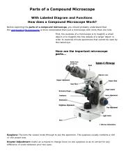
Compound Microscope Parts, Functions, and Labeled Diagram ...
Compound Microscope: Definition, Diagram, Parts, Uses, Working ... - BYJUS A compound microscope is defined as A microscope with a high resolution and uses two sets of lenses providing a 2-dimensional image of the sample. The term compound refers to the usage of more than one lens in the microscope. Also, the compound microscope is one of the types of optical microscopes.

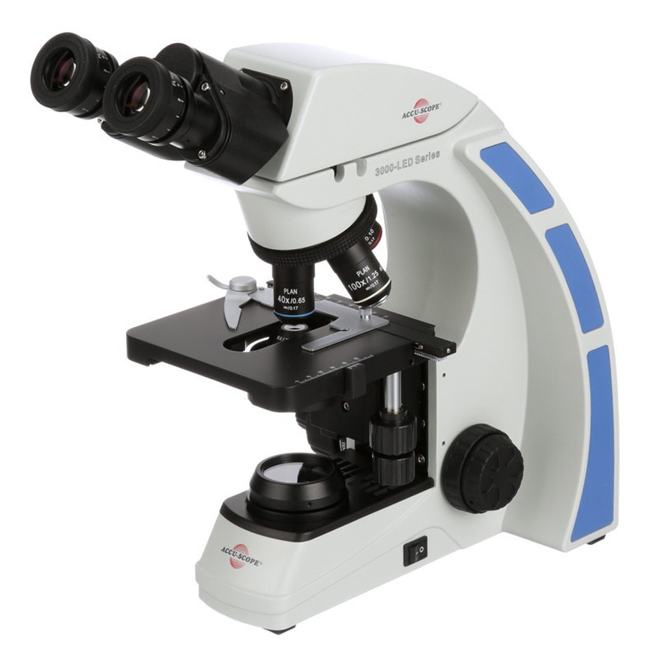
AmScope 40X-2500X LED Lab Binocular Compound ...
Compound Microscope Labeled Diagram | Quizlet Compound Microscope Labeled + − Flashcards Learn Test Match Created by meganplocher734 Terms in this set (14) Eyepiece/Ocular lens Contains the ocular lens Body tube A hollow cylinder that holds the eyepiece. Arm Part that supports the microscope. Stage Supports the slide or specimen Coarse adjustment Knob

List: Parts of a Microscope and their Function | Pathwooded
Imaging Subcellular Structures in the Living Zebrafish Embryo The relative ease with which these subcellular structures can be genetically labeled by fluorescent proteins and the use of light microscopy techniques to image them is ... it is also a fluorescent compound itself. Therefore, Phenol Red could obscure visualizing FPs if imaging is planned before 24 hr post-fertilization (hpf) as it may not be ...

Compound microscope – INNOVATIVE CREATIONS

A Study of the Microscope and its Functions With a Labeled ...

Monday, 12 November 2018Monday, 12 November ppt download

Compound microscope labeling Diagram | Quizlet
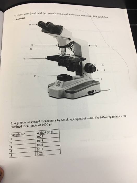
Solved Identify and label the parts of a compound microscope ...

16 Basic Parts of Microscope, Function, Names & Labeled Diagram
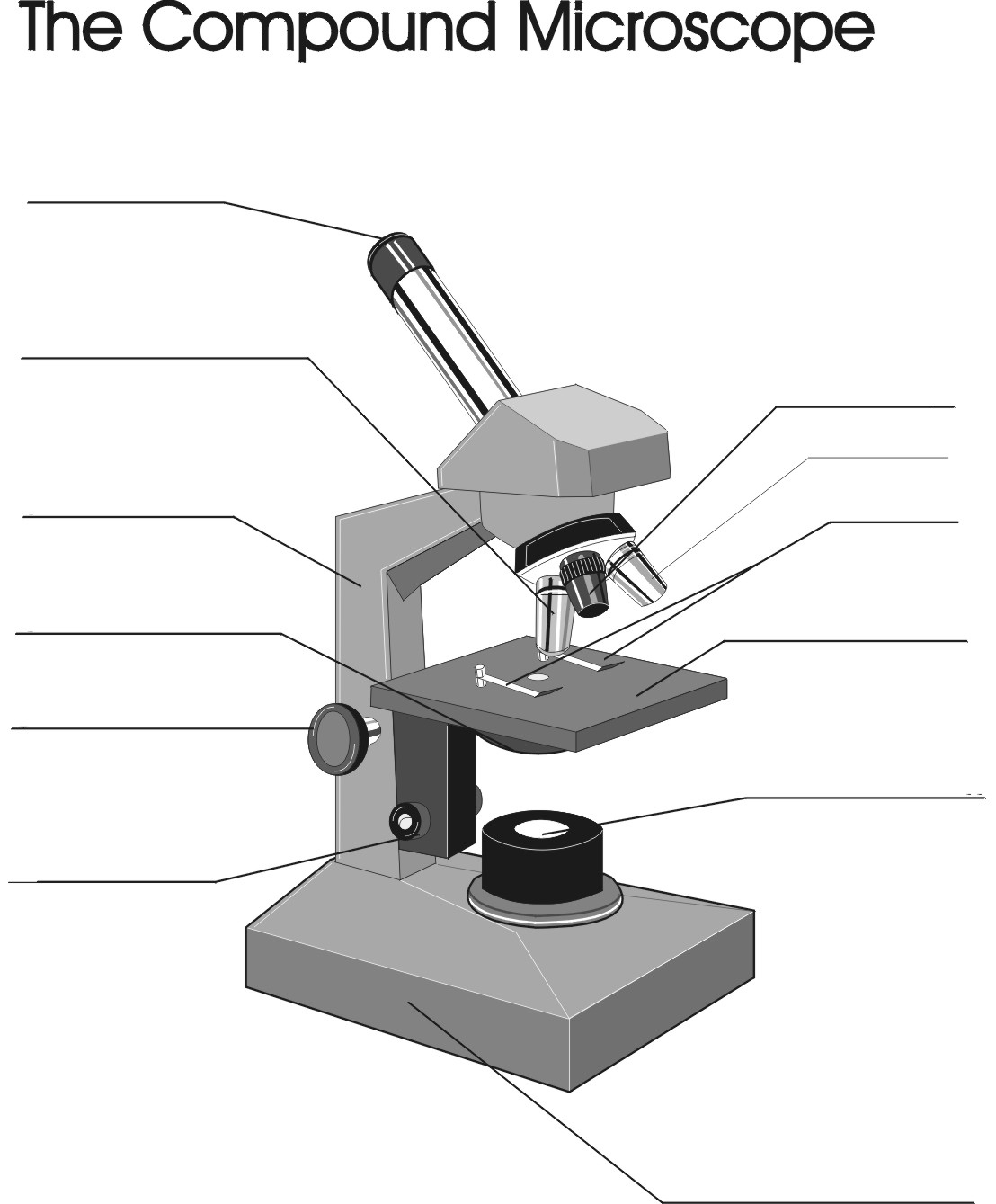
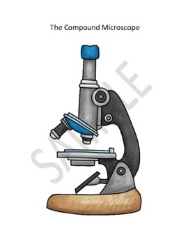
Remix of "The Compound Microscope"

Compound Microscope- Definition, Labeled Diagram, Principle ...

Microscope, Microscope Parts, Labeled Diagram, and Functions

Compound Microscope Parts Made Easy

Diagram of a Compound Microscope

File:Microscope diagram.png - Wikimedia Commons
Exercise 1: Using a Compound Microscope | SpringerLink

Compound Microscope Parts, Functions, and Labeled Diagram ...

The Microscope

Label the Microscope Diagram | Download Scientific Diagram
Compound Microscope Parts, Functions, and Labeled Diagram ...
Using A Compound Microscope Teaching Resources | TpT
MICROBIO 16 Parts of a Compound Microscope with Diagram and ...

Below is a photo of a compound light microscope with labels ...

This is a common compound microscope Label its parts class 11 ...

Binocular Compound Light Microscope Diagram Quiz

How to draw compound of Microscope easily - step by step
Microscope Diagram Labeled, Unlabeled and Blank | Parts of a ...

Microscope Maintenance Tips | Science supplies, Microscope ...

Types of Microscopes: Definition, Working Principle, Diagram ...

Leica Upright Compound Light Microscope Diagram Diagram | Quizlet
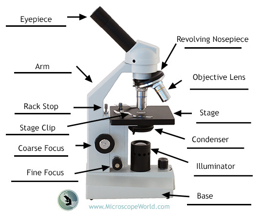
Labeling the Parts of the Microscope | Microscope World Resources

Parts of a Microscope - SmartSchool Systems
Best Compound Microscope – A Comprehensive Guide - Microscope ...

This is a common compound microscope. Label its parts from A ...


Komentar
Posting Komentar